Nuclear isolation protocol
This nuclear isolation protocol uses hypotonic lysis and Dounce homogenisation for nuclear fractionation of adherent mammalian cells. You separate nuclear and cytoplasmic fractions by differential centrifugation. The cytosolic supernatant and nuclear pellet can be extracted directly into SDS sample buffer for western blotting (e.g. transcription factor translocation, signalling intermediates). For a faster Triton-based soluble vs insoluble prep suited to F-actin:G-actin ratios, see our F:G actin ratio protocol.
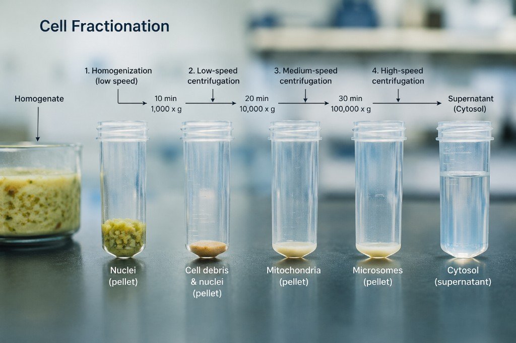
Nuclear fractionation overview
Cells are swollen in hypotonic buffer so the plasma membrane ruptures with minimal mechanical force. A loose Dounce pestle releases nuclei without over-shearing chromatin. Incomplete lysis is checked by phase-contrast microscopy before fractions are separated by centrifugation. Work on ice throughout; pre-chill centrifuges, tubes, homogeniser, and buffers.
- Scale: use T75 flasks or larger — smaller dishes rarely yield enough material.
- Timing: allow ~45–90 min from harvest to samples in SDS buffer (excluding stimulation).
- Yield check: a compact white nuclear pellet should be visible after the wash spin; the cytosolic supernatant should be clear, not cloudy with debris.
Before you start
- Cold PBS, wash buffer, and freshly made hypotonic nuclei lysis buffer (with protease inhibitors).
- Trypsin (or your usual detachment reagent) and ice-cold 10% FCS to quench.
- 15 ml conical tubes, 1.5 ml Eppendorf tubes, serological pipettes.
- 2 ml glass Dounce homogeniser with loose (type B) pestle.
- Phase-contrast microscope and tissue-culture plate for smear checks.
- Refrigerated microcentrifuge; note your rpm-to-g conversion for reproducibility.
- 1× reducing SDS sample buffer; β-mercaptoethanol or DTT per your lab standard.
Procedure
- Culture and stimulation. Plate cells in tissue-culture flasks (no smaller than T75) and treat with the agonist or condition under investigation. Harvest at the desired time point.
- Cold PBS wash. Aspirate medium and wash monolayers twice with ice-cold PBS to remove serum and salts. Work quickly so cells do not warm on the bench.
- Harvest. Detach with trypsin (or equivalent). Neutralise with an equal or greater volume of ice-cold 10% FCS in PBS or growth medium. Transfer to a 15 ml tube and pellet cells at 1400 rpm (~300–400 g in a typical swinging-bucket rotor; adjust for your centrifuge) for 5 min at 4 °C.
- Discard supernatant.
- Wash 1. Resuspend pellet in 10 ml ice-cold wash buffer. Mix gently by inversion.
- Pellet again at 1400 rpm, 5 min, 4 °C.
- Discard supernatant.
- Hypotonic swelling. Resuspend cells in 0.75–1.0 ml ice-cold hypotonic nuclei lysis buffer and transfer to a 1.5 ml Eppendorf on ice. Incubate on ice for 5–10 min so cells swell before homogenisation.
- Homogenise. Transfer the suspension to a 2 ml glass Dounce homogeniser on ice. Homogenise with 10 slow, steady strokes of the loose pestle (draw pestle up without twisting; avoid foaming).
-
Microscopy checkpoint. Remove 5 µl of homogenate and smear onto a
well of a tissue-culture plate. Examine by phase contrast at high magnification:
- Nuclei — small, dark, refractile ovals.
- Unlysed cells — larger, brighter, rounded bodies.
- If many intact cells remain, repeat homogenisation (10 strokes per round) and re-check. Avoid excessive passes, which can shear nuclei and contaminate the cytosolic fraction with nuclear proteins.
- Separate nuclei. Spin homogenate at 3000–4000 rpm in a standard microcentrifuge (~2000–3000 g; confirm for your rotor) for 3 min at 4 °C. A compact pellet of nuclei should form.
- Collect cytosol. Carefully transfer the supernatant (cytoplasmic fraction) to a fresh cold 1.5 ml Eppendorf on ice. Do not disturb the nuclear pellet.
- Wash nuclei. Gently resuspend the nuclear pellet in 1 ml ice-cold wash buffer (pipette along tube wall; avoid vigorous vortexing).
- Spin again: 4000 rpm, 3 min, 4 °C.
- Remove wash buffer. Aspirate or pipette off supernatant completely. A small volume of residual liquid is acceptable; carry-over dilutes SDS buffer minimally.
- SDS extraction — nuclei. Resuspend the nuclear pellet in 1× reducing SDS sample buffer. Use a similar volume to the cytosolic fraction for comparable loading. As soon as SDS contacts the pellet, pipette rapidly up and down — the extract becomes very viscous (chromatin) and can form a gel-like clump if left static. Repeat pipetting until viscosity drops and the sample is homogeneous.
- SDS extraction — cytosol. Add an equal volume of 1× reducing SDS sample buffer to the cytoplasmic supernatant (or adjust to match nuclear volume). Mix thoroughly.
- Denature for SDS-PAGE. Heat both fractions at 95 °C for 5 min, vortex briefly, spin down, and load on gel. Store aliquots at −20 °C if not run immediately.
Tips and troubleshooting
- Low nuclear yield: increase flask area, ensure complete trypsinisation, and confirm homogenisation — under-lysis leaves nuclei trapped in intact cells.
- Cytoplasmic contamination of nuclei: extend the wash-buffer spin (step 15–16); optional second wash with 0.5 ml wash buffer if background bands persist.
- Nuclear proteins in cytosol: usually over-homogenisation or pellet disturbance when removing supernatant — use fresh pipette tips and leave a few µl above the pellet if needed.
- Viscous nuclear SDS sample: normal; add buffer in steps, shear with repeated pipetting, then heat before loading.
- Protease inhibitors: include AEBSF or a cocktail in hypotonic lysis buffer; add fresh each preparation.
Stock solutions
Prepare from analytical-grade reagents; filter or autoclave where your lab requires sterility for storage stocks.
| Stock | Storage |
|---|---|
| 1 M NaCl | Room temperature |
| 1 M KCl | Room temperature |
| 1 M HEPES pH 7.9 | Room temperature |
| 1 M MgCl2 | Room temperature |
| 1 M DTT — prepare 10 ml, aliquot | −20 °C |
| 1 M EDTA — adjust to pH 8.0 with NaOH (may not dissolve until alkaline) | Room temperature |
| 10% NP-40 (or IGEPAL CA-630) | Room temperature |
| 100 mM AEBSF (or protease inhibitor cocktail per manufacturer) | −20 °C or fresh |
Wash buffer (50 ml)
Final concentrations: 10 mM HEPES pH 7.9, 10 mM KCl, 1.5 mM MgCl2. Chill before use.
| Component | Volume |
|---|---|
| 1 M HEPES pH 7.9 | 0.5 ml |
| 1 M KCl | 0.5 ml |
| 1 M MgCl2 | 75 µl |
| Ultrapure H2O | to 50 ml (48.925 ml) |
Hypotonic nuclei lysis buffer (10 ml)
Make fresh each experiment. Final concentrations: 10 mM HEPES, 10 mM KCl, 1.5 mM MgCl2, 1% NP-40, 0.5 mM AEBSF (or equivalent inhibitor cocktail). Keep on ice.
| Component | Volume |
|---|---|
| 10% NP-40 stock | 100 µl |
| 100 mM AEBSF (or protease inhibitor cocktail) | 50 µl |
| 1 M HEPES pH 7.9 | 100 µl |
| 1 M KCl | 100 µl |
| 1 M MgCl2 | 15 µl |
| Ultrapure H2O | 9.635 ml |