Western blot lysis buffer & blocking buffers
How to quantify a western blot: After transfer and imaging, use our western blot quantification tool to draw ROIs, subtract background, and export adjusted integrated density to CSV.
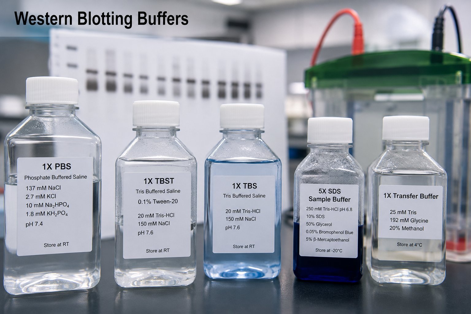
If you are looking for a western blot lysis buffer or a lysis buffer for western blot, you will often see RIPA first. Many researchers new to western blotting land on a recipe for RIPA buffer. That choice is very often not the most efficient, the cheapest, or the simplest to make for immunoblotting. The name is the clue: RIPA stands for radioimmunoprecipitation buffer. It is formulated to be gentle enough for immunoprecipitation assays so antibody–antigen interactions are not disrupted. As a result it is relatively poor at solubilising cytoskeletal proteins and extracting nuclear material compared with a more denaturing SDS-based lysis mix.
SDS lysis buffer for western blotting
For western blotting, a much more efficient approach is a lysis buffer with a high concentration of SDS. The recipe below is typical of what many labs use when the goal is maximum protein solubilisation for SDS-PAGE and immunoblotting, not gentle IP conditions.
Protease and phosphatase inhibitors: unlike RIPA workflows, you usually do not need expensive inhibitor cocktails here. The high SDS concentration partially denatures proteases and phosphatases and blocks much of their activity. Always follow your own lab safety rules for β-mercaptoethanol and SDS.
SDS lysis buffer (50 ml)
| Reagent | Amount (50 ml) |
|---|---|
| Glycerol | 10 ml |
| 500 mM Tris, pH 6.8 | 5 ml |
| 10% (w/v) SDS | 10 ml |
| β-Mercaptoethanol | 125 µl |
| Bromophenol blue | Add powder until the mix is a reasonable blue (tracking dye for loading) |
Mix components in the order that works best in your lab (often Tris and SDS first, then glycerol and β-mercaptoethanol, with bromophenol blue last). Store aliquots according to your SOP; many labs prepare fresh or short-term stocks because of reducing agent stability. Wear appropriate PPE when handling SDS and β-mercaptoethanol.
This buffer is closely related to standard SDS sample-loading formulations and pairs well with protocols that separate cytosolic and cytoskeletal fractions using SDS, such as the F-actin ratio method in our protocols collection.
Blocking buffer for cleaner blots
Another helpful step for clean western blots is a blocking buffer that combines milk, BSA, and a low level of detergent. The recipe below (“Super Western blot blocking buffer”) is heated and filtered so particulate matter from the milk does not end up on the membrane.
Super Western blot blocking buffer
| Component | Final concentration |
|---|---|
| Tris-buffered saline (TBS) | 1× (base solution) |
| Non-fat dried milk powder | 2.5% (w/v) |
| BSA | 2.5% (w/v) |
| Triton X-100 | 0.2% (v/v) |
- Combine ingredients in 1× TBS. Microwave until hand-hot (about 50–60 °C) to help the milk powder dissolve.
- Stir with a magnetic stirrer for 10 minutes.
- Adjust pH to 7.6. Milk powder lowers the pH of TBS, so always re-check and re-adjust pH after adding milk.
- Centrifuge at 3,000–4,000 × g for 30 minutes. A soft pellet of sludge will form; remove this material so it does not transfer onto the blot.
- Filter the supernatant through a 0.45 µm filter. Use the clarified buffer for blocking.
Use this buffer in place of plain milk or BSA blocking where background or speckling from undissolved milk solids has been a problem. As with any blocking formulation, antibody compatibility (especially phospho-specific antibodies) should be checked in your system.
Summary
For western blotting, favour an SDS-rich lysis buffer when you need efficient extraction rather than IP-grade gentleness. Pair it with a filtered milk/BSA blocking buffer when you want to reduce membrane artefacts from milk particulates. Adjust recipes to your sample type, antibody, and institutional health and safety requirements.